Abstract
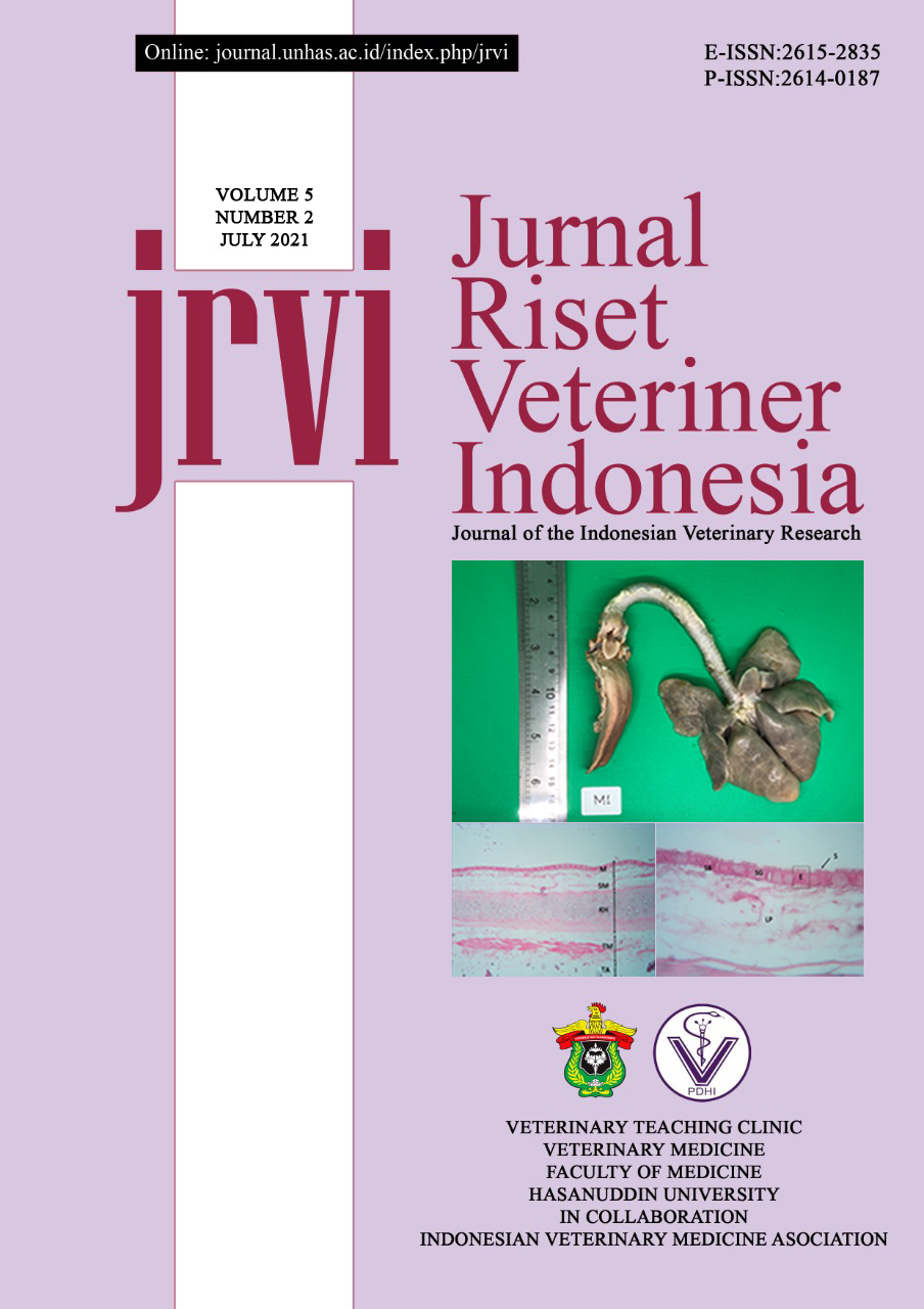
The common palm civet (Paradoxurus hermaphroditus) is one of the wild animals that can be found across Java, Sumatra, Kalimantan, Sabah, Sarawak, India, and Sri Lanka. Civet is considered as a nocturnal and arboreal animal. This study aims to determine the anatomical morphology of the civet’s trachea, which information is still limited. The trachea of three adult civets of different sexes obtained from Yogyakarta and Lampung were used in this study. Samples were collected, diffused, examined macroscopically, and microscopically processed to produce paraffin blocks. Paraffin blocks were sliced of thickness 4µm using a microtome. Samples were stained with hematoxylin-eosin. The staining results were then described to determine the tracheal profile of the civet’s trachea. Histologically, trachea has several layers successively from the inside out are the mucosa, submucosa, hyaline cartilage layer, and tunica adventitia. The mucosal layer consists of the epithelium, lamina propria, and the invisible muscularis mucosa. The epithelium portion of the trachea is a ciliated stratified pseudo columnar containing goblet and basal cells. Column cells are oval with a dark cell nucleus at the basal area. The lamina propria part of the civet’s trachea consists of connective tissue consisting of collagen, elastin, and reticular fibers. There are seromucous glands between the lamina propria and submucosa. The tracheal musculus is located on the external side of the hyaline cartilage.
Keywords: Common Palm Civet, Hematoxylin Eosin, Histology, Morphology, Trachea

This work is licensed under a Creative Commons Attribution-NonCommercial 4.0 International License.

