Abstract
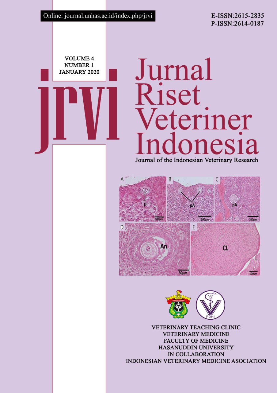
Female reproductive system showing the fastest signs of aging. The ovarian aging characterized by a decrease in follicular development. Stem cells are undifferentiated cells and can form a variety of different cells as the foundation of tissues and organs. Previous studies reported that Bone Marrow Mesenchymal Stem Cells (BM-MSCs) transplantation can restore follicular development in damaged ovarian rats. This study aimed to analyze the number of follicular development in aged rats and to analyze the capability of human Umbilical Cord Mesenchymal Stem Cells (hUC-MSCs) to improving follicular development in aged rats. This study used 3 mature rats (4 months old), and 9 nine aged rats (22-24 months old), Spraque Dawley (SD) strain. They were divided into four groups. The first and the second group was mature rats and aged rats without injection. The third and the fourth group was aged rats injected hUC-MSCs dose 106 cells/kgBW and hUC-MSCs dose 107 cells/kgBW. The injection carried out 4 times at 3-month intervals. The parameters observed were follicular development and homing image of hUC-MSCs in ovarian tissue. The results showed that the number of follicular developments in aged rats 22-24 months decreased significantly compared to mature rats 4 months old. Injection of hUC-MSCs at dose 106 cells/kgBW and 107 cells/kgBW did not increase follicular development in aged rats. hUC-MSCs did not found in ovarian tissue. It could be concluded that aged rats 22-24 months old no longer productive indicated from the number of follicular developments and corpus luteum decreased. The injection of hUC-MSCs intravenously did not indicate an improvement of follicular development in aged rats 22-24 months old.
References
Acuna E et al. 2009. Increases in norepinephrine release and ovarian cyst formation during ageing in the rat. Reproductive Biology and Endocrinology, 7:64
Batsali AK et al. 2013. Mesenchymal Stem cells Derived from Wharton's Jelly of the Umbilical Cord: Biological Properties and Emerging Clinical Applications. Curr Stem Cell Res Ther, 8(2):144–155
Cho PS et al. 2008. Immunogenicity of umbilical cord tissue–derived cells. Blood, 111(1): 430–43
Conconi MT et al. 2011. Phenotype and Differentiation Potential of Stromal Populations Obtained from Various Zones of Human Umbilical Cord: An Overview. The Open Tissue Engineering and Regenerative Medicine Journal, 4:6–20
Edessy M et al. 2014. Stem cells Transplantation in Premature Ovarian Failure. WJMS, 10(1): 12-16
Garg N dan Sinclair DA. 2015. Oogonial Stem cells as A Model to Study Age-Associated Infertility in Women. Reprod Fertil Dev, 27(6): 969-974
Gonçalves JTT et al. 2016. Overweight and obesity and factors associated with menopause. Ciênc. Saúde Colet, 21(4)
Kothiyal P dan Sharma M. 2013. Postmenopausal quality of life and associated factors- A Review. Journal of Scientific and Innovative Research, 2(4): 814-823
Niccoli T dan Partridge L. Ageing as a risk factor for disease. Curr Biol, 11:R741-52
Pedersen T dan Peters H. 1968. Proposal for a classification of oocytes and follicles in the mouse ovary. J Reprod Fertil. 17(3):555-557
Pérez-López Fr et al. 2009. Cardiovascular risk in menopausal women and prevalent related co-morbid conditions: facing the post-women’s health initiative era. Fertil. Steril. 92:1171–1186
Sensebé L dan Fleury-Cappellesso S. 2013. Biodistribution of Mesenchymal Stem / Stromal Cells in a Preclinical Setting. Stem Cell International, 678063:1-5.
Vladislav V et al. 2014. Stem cells as New Agents for The Treatment of Infertility: Current and Future Perspectives and Challenges. Biomed Research International, 507234:1-8
Zhu SF et al. 2015. Human Umbilical Cord Mesenchymal Stem Cell Transplantation Restores Damaged Ovaries. J Cell Mol Med, 19(9): 2108-2117

