
Keywords
Red spinach
Kidney
Anatomy change
Histology change
How to Cite
Abstract
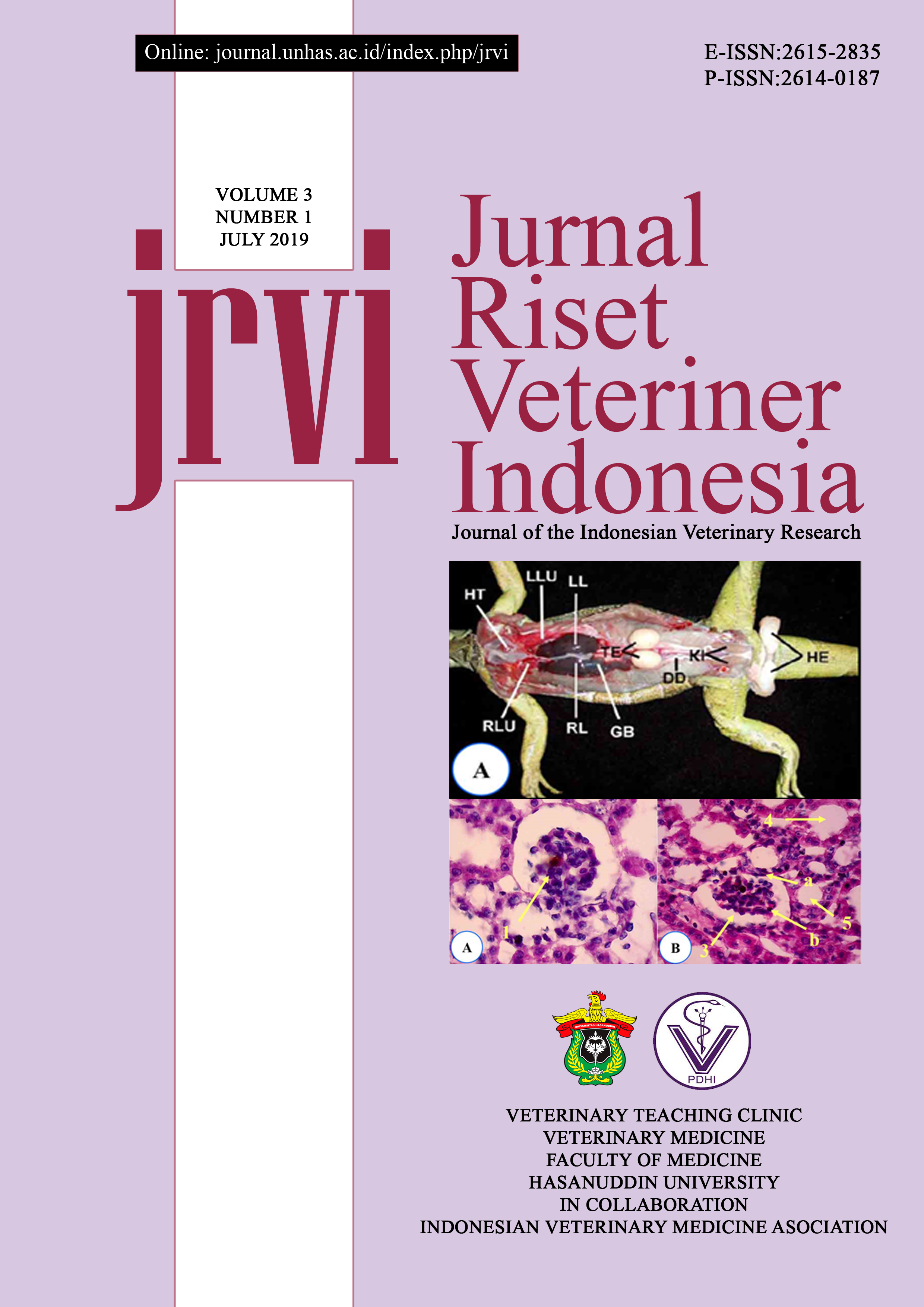
Background and Objective: Iguana breeders usually used red spinach as a feed for iguanas. However, several studies have indicated that red spinach contains oxalate and purine which are harmful if consumed at a certain level because they can interfere with kidney function. This study aimed to investigate the change of anatomy and histology of green iguana (Iguana iguana) kidney after feeding with red spinach. Materials and methods: Twelve iguanas were used in this study and were divided into four groups in accordance to the dose level of red spinach (100%, 75%, 50% and 25%, namely group I to IV, respectively). The red spinach was given for 30 days through the nasogastric intubation. The effect of red spinach administration was observed through the anatomy and histological changes. The results were statistically analyzed with One Way ANOVA with p<0.05 was considered significant, and Post Hoc Test. The histopathology change of the kidney was analyzed descriptively. Results: The administration of red spinach in iguanas caused some anatomy changes of the kidney; such as the enlargement of iguana’s kidney. The histological findings included the enlargement of the glomerulus, capsular space constriction, hydropic degeneration, tubular dilation, necrosis, and formation of connective tissue (fibrosis), uric acid crystal sediment (gout), oxalate crystal, and lymphocyte infiltration. Among four different dose levels of red spinach, 100% of red spinach caused the highest damage to the iguana’s kidney. The significant change grew as the increasing dose of red spinach that was given to the green iguanas’ bodies. Conclusion: Red spinach caused changes in anatomy structure as well as kidney histology of green iguanas. Severe damages occurred in the treatment group III, moderate damage occurredin the treatment group II, and light damage occurred in the treatment group I.
Keywords: Green iguana, Red spinach, Kidney, Anatomy Change, Histology Change